Dr. Karoly: Plant Evolution
Preparing for data collection: Microscopes and Ocular Micrometer Calibration
How-to videos for a Dissecting (Stereo) Microscope, Compound Microscope, and Ocular Micrometer Calibration
(closed captioning is available for each video using the CC control in the video window)
|
Dissecting Microscope (2:57') |
Compound Microscope (11:17') |
Ocular Micrometer (7:00') |
Spore and Pollen Videos
Videos showing the location for spores and pollen grains and the method for preparing your wet mount slides.
(closed captioning is available for each video using the CC control in the video window)
MOSS video (3:50') |
FERN video (2:30') |
CONIFER video (4:30') |
FLOWERING PLANT video (2:30') |
Spore and Pollen Images
Below are observational data that provide information about the composition of spores and pollen grains. They images will give you data regarding the number of nuclei they contain and the chemical composition of their outer wall.
Number of nuclei in spores and pollen grains
DAPI binds to DNA and thus allows the location of nuclei to be determined, as they will stain a bright blue.
Spores of a moss and a fern and pollen from a conifer and a flowering plant were each stained with DAPI and visualized under UV fluoresence.
Look at the spores and pollen and look for brightly colored (blue) structures inside.
Do spores and pollen have the same number of nuclei?
clicking on an image will open a new window with a larger version of the image (approx. 800 x 600).
MOSS |
FERN |
CONIFER |
FLOWERING PLANT |
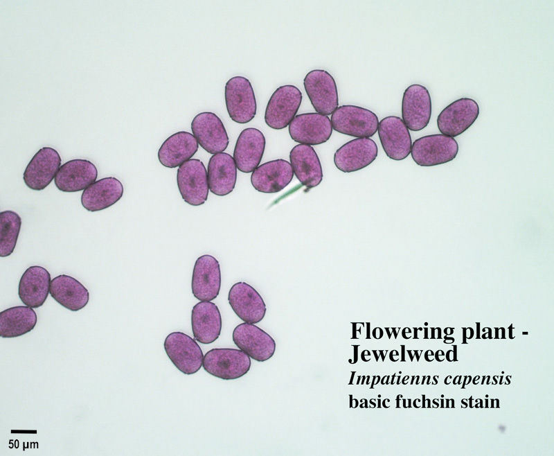
Staining by basic fuchsin in spores and pollen grains
Basic fuchsin is the stain component of Calberla's Pollen Stain which is used to differentiate pollen grains from other materials in geology (palynology) and in meterological sampling for allergens (aerobiology). Basic fuchsin positively stains the outer layer of the pollen grain wall (the exine) a dark pink to red color.
Spores of a moss and a fern and pollen from a conifer and a flowering plant were each stained with basic fuchsin and visualized under visible-light illumination.
We expect pollen to stain dark red because this stain is used as a way to identify pollen.
Do the spores also stain the dark red color seen in the pollen?
clicking on an image will open a new window with a larger version of the image (approx. 800 x 600).
MOSS |
FERN |
CONIFER |
FLOWERING PLANT |
Pollen and Spore Image Collections on the Web
PalDat - Palynological Database
PalDat is the world's largest pollen database. It is organized by taxon and provides descriptions and images of pollen and the associated flowers/inflorescences. It is operated by members of the Division of Structural and Functional Botany (SFB) at the University of Vienna and supervised by AutPal, the Society for the Promotion of Palynological Research in Austria.
Pollen Atlas of bat-pollinated plants
From the New York Botanical Garden, this site features SEM images of pollen collected from the diverse species of flowering plants that are pollinated by bats in Central French Guiana.