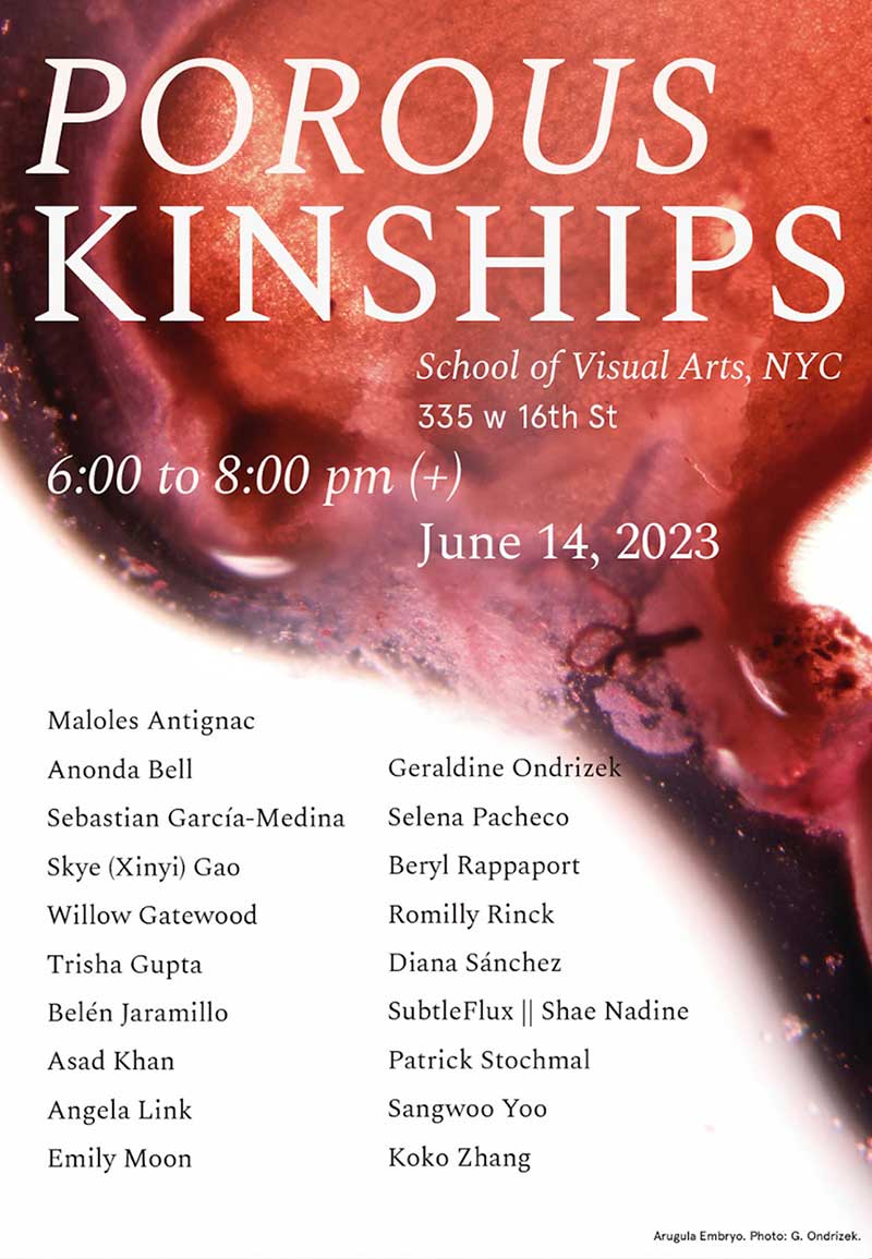
Germination/Gestation
I have been making works about in-vitro fertilization, early stage cell development, and the dynamics of cellular division over the past two years. I have found that the ability for cells to divide, thrive and make human, animal and plant embryos is nothing short of magic. Both plant and animal embryos have epithelial cells, which are amazingly elastic, rapid growing cell tissues that are in contact with the interior and exterior of organisms. Epithelial cells create a protective layer on a plant’s roots, and are responsible for human neural tube development.
With these cells in mind during this SVA Bio Art Residency, I planted seeds. On May 24 and 27 I planted seeds and set up germination tubes. I was astonished at the quick germination and growth and photographed these every day. Once we learned to use the microscopes, ( ) I was able to harvest tiny plants and seed embryos for observation. These are the photos of epithelial cells that resulted.
In observing seed growth, I witnessed properties of seeds that are nothing short of superpowers. In a dry state they can store their energy for years and then suddenly release it for germination when environmental conditions are favorable. Researchers discovered that when the seeds came into contact with water, energy metabolism was established in a matter of minutes, and the plant cells' "power stations", known as mitochondria, activated their respiration.
Seed embryos live off the seed sacks and water until the epithelial cells are formed and nutrients from water and the ground are able to be absorbed. Plant epithelial cells are mainly found in the epidermis which covers the vascular cylinder of the inner root. (See images of the arugula root and sunflower root, in which the dark inner mass is the center). Epithelial cells are fundamental for nutrients to filter into the vascular center and prevent them from flowing back out.
In human and animal embryonic development, Epithelial-mesenchymal transition (EMT) occurs, which from the neural tube responsible for the vascular network, the spine, nervous system, and skin are grown. These are also known as stem cells.
EMT and Mesenchymal-epithelial transition (MET) are evolutionarily conserved processes that are used throughout embryonic development to drive tissue morphogenesis. During adult life, EMT is activated to close wounds after injury, but also can be used by cancers to promote metastasis. This dynamic is astonishing, and the core reason cancers have the ability to live and thrive.¹
The images I photographed in this residency are of the epithelial cells of plants. Because plant cells are colorless, I injected the plants with Safranine, an azo dye commonly used for plant microscopy, which turns the cell walls a red/ orange color or Toluidine blue staining that helps see the water flow through the roots. I took multiple photographs so that the entire length of the root and plant could be understood as it recently emerged from the embryonic state. Observing the forms and systems closely, I found how the root looks like a vein, which in fact it is. Although I had hoped to make more images of plant embryos, the Arugula embryo was the closest I could get.
List of works:
1. Leaf to Root-
2. Basil leaf, stem, seed, root
3. Slide Mounts
4. Pea , Leaf, Root
5. Sun Flower Root
6. Arugula First Root
7. German Winter Thyme Seed To First Root
8. Arugula Embryo
Bio Art Artist in Residence Summer 2023
From the Laboratory to the Studio: Interdisciplinary Practices in Bio Art
The Bio Art summer interdisciplinary residency takes place in the new SVA Bio Art Laboratory, located in the heart of New York City’s Chelsea gallery district. The SVA Bio Art Lab houses microscopes for photo and video, skeletons and specimen collections, a herbarium and an aquarium as well as a library. Each resident is awarded a private studio space. The residency culminates in a public exhibition.
The Residency is led by artist Suzanne Anker, chair of the SVA BFA Fine Arts Department; and Joseph DeGiorgis, marine biologist.
¹ Epithelial–mesenchymal transition was first recognized as a feature of embryogenesis by Betty Hay in the 1980s. EMT, and its reverse process, MET (mesenchymal-epithelial transition) are critical for development of many tissues and organs in the developing embryo, and numerous embryonic events such as gastrulation, neural crest formation, heart valve formation, secondary palate development, and myogenesis.[3] Epithelial and mesenchymal cells differ in phenotype as well as function, though both share inherent plasticity. Epithelial cells are closely connected to each other by tight junctions, gap junctions and adheres junctions. They have an apico-basal polarity, polarization of the actin cytoskeleton, and are bound by a basal lamina at their basal surface. Mesenchymal cells, on the other hand, lack this polarization, have a spindle-shaped morphology and interact with each other only through focal points. Epithelial cells express high levels of E-cadherin, whereas mesenchymal cells express those of N-cadherin, fibronectin and vimentin. Thus, EMT entails profound morphological and phenotypic changes to a cell.


