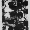
De Viscerum Structura
WORKS IN THE EXHIBITION
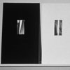
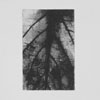
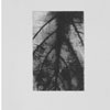
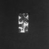
OVUM
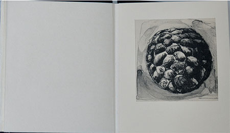
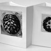
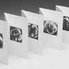
13 Magnified cell lithographs on mulberry paper 7" x 8" when closed, and 8' long open. Each image is 3" x 3"
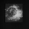
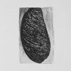
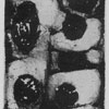
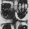
The central image is 4" x 4". This thirteen-print series documents the development of the ovum through the blastocyst stage, in which each cell is identified as a particular part of the human anatomy. The original cells were magnified 900 times with an electron microscope. The images were scanned, enlarged, and altered on a computer, then transferred on to lithographic plates.
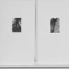
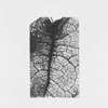
CELLULAR TISSUE
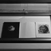
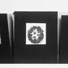
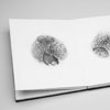
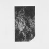
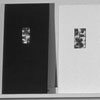
10 Magnified plant tissue lithographs on mulberry paper, ink, bound in black folio 10” x 18”
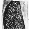
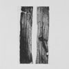
CHROMOSOMES
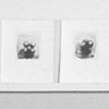
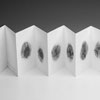
8 cortical cells lithographs on mulberry paper, bound in black folio
3” x 4”
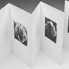
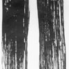
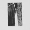
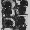
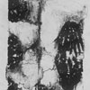
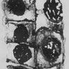
This book of eight prints shows the longitudinal files of incipient cortical cells, which are still actively dividing. Each image crops into the tissue to focus on the various stages of mitosis and the threads of the chromosomes. The images were scanned, enlarged, and altered on a computer, then transferred lithographic plates. Two of the editions are unbound; one is bound as an accordion so that the book unfolds and stands on its own.
De Viscerum Structura - Latin, meaning the structure of flesh
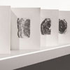
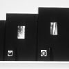
An installation of ten books. In this work I have focused on the magnification of organic forms and our cultural preoccupation with inspection. With the invention of the microscope in the Seventeenth Century, the interior of nature, once closed off to direct perception, was made accessible. This development spurred debates about realism and instrument-mediated knowledge, which are echoed today. Most recently the use of magnification has vastly expanded our knowledge of human reproduction, genetic coding, and fetal tissue. These new frontiers have changed our understanding of and ability to deal with disease and reproduction.
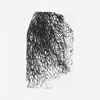
My preoccupation with inspection and collection naturally led me to an investigation of the magnification of organic matter. I began my first piece, Tissues, by directly scanning plant tissue into the computer and magnifying it ten to twenty times. Through this process I found that the tissues became so abstract they could be read as many types of organic matter. Simultaneously I researched the images made with an electron microscope. I accessed several resources of images of plant tissue and human fetal cell growth. The visual simplicity of these abstract calligraphic marks and the potent content of fetal cell tissue were of particular interest. The framing and diagrammatic display of these images in medical journals and scientific texts gave me a better understanding of the process of division and formation. This didactic format in juxtaposition with the actual content of each cell further intrigued me: each cell carries an entire map of a human being, representing pure potential.
In the book Ovum I focused on making images of the blastocyst stage, in which the cells are identified as a specific part of the body and divide, creating the mid-line or spinal column of the body. I then began looking at images of chromosomes actively dividing in cellular tissue. Each image in the book Chromosomes focuses on the various stages of mitosis.
The books were produced in the summer and fall of 1998, while on a junior faculty paid leave from Reed College. The project was supported by the Stillman-Drake fund and the Levine Funds. The prints were produced at Mahaffey Fine Art in Portland. The books were constructed and bound while I was an artist in residence at the Women's Studio Workshop in New York.
