Screening of Cellular
Fourth Annual Feminist Art History Conference,
American University and the National Museum of Women in the Arts, Washington, DC
2013
Cellular 2009
12 min.
Digital Film made with Atonics Micro fire digital cameras mounted on Olympus stereomicroscopes
Sound made with AMF, atomic force microscope
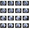
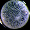
Cellular is a film of a blastopore, or a multiple-cell embryo. More than 200 hours of still images of each embryo were made with Atonics Micro fire digital cameras mounted on Olympus stereomicroscopes and made into a film using Astor IIDC imaging software. My colleague at Reed College, Steve Black, Professor of Developmental Biology and Zoology, provided me with a lab space to make the films of the blastopore. I worked with his lab assistant Allison Egar to make a film of development which was used for the still images. To make my film, I edited 10 film sections to overlap and repeat the phases of development just before a recognizable body is evident. Each segment represents the different eggs in the process of development. Each is edited prior to full gestation, creating a loop of endless potential. The film Cellular is overlaid with the recording of the Sound of Cells Dividing.
Since 2003, nanotechnology and the AMF (atomic force microscope) pioneered by James Giminski, Professor of Chemistry at UCLA, and Andrew Pelling Associate Professor of Biophysics at the University of Ottawa, have enabled researchers to also touch a cell. Like the blind reading braille or a finger[finger is more commonly used] touching the pulse, the needle of the AMF can touch cells which are less than half the diameter of a human hair. This touch can also record the sound the cell emits when dividing or dying. Through touching and hearing rather than only seeing, an entirely new knowledge base has emerged, one that artists have known, that of the multi-sensory knowledge. What we can now listen to within a cell has revolutionized our knowledge of cellular gestation, division, and the relationships within interior architecture of the body.
The sounds one hears in the film Cellular is five tracks of cells, some healthy, some damaged, dividing and dying. I accessed these recordings from James Gimzewski, Professor of Chemistry at UCLA and Andrew Pelling Associate Professor of Biophysics at the University of Ottawa. The recorded cells vibrate at nanoscale and the vibrations are then amplified, bringing the sounds into the range of human hearing. The new area of study is called "sonocrology”. The main tool for learning about cell sounds is the atomic force microscope (AFM). Instead of using optics to show an image, the AFM uses a very fine tip to feel a cell in the same way a needle was used to feel the pattern of vibrations pressed into vinyl records. Gimzewski and Pelling have found that cells with cancer or other diseases emit very low and strained frequencies, while healthy cells emit a pleasant sounds. Sonocytology has proven to be a noninvasive way to detect disease. In 2007, Gimzenwski used nanotechnology to demonstrate that metastatic cancer cells are softer than healthy cells. The study represented one of the first times researchers have been able to take living cells from human cancer patients and use nanotechnology to determine, through touch, which were cancerous.
Special Thanks; Professor Steve Black, Allison Edgar, and Judith Levine for the raw film clips.

